Dilated cardiomyopathy in Dobermanns: echocardiography
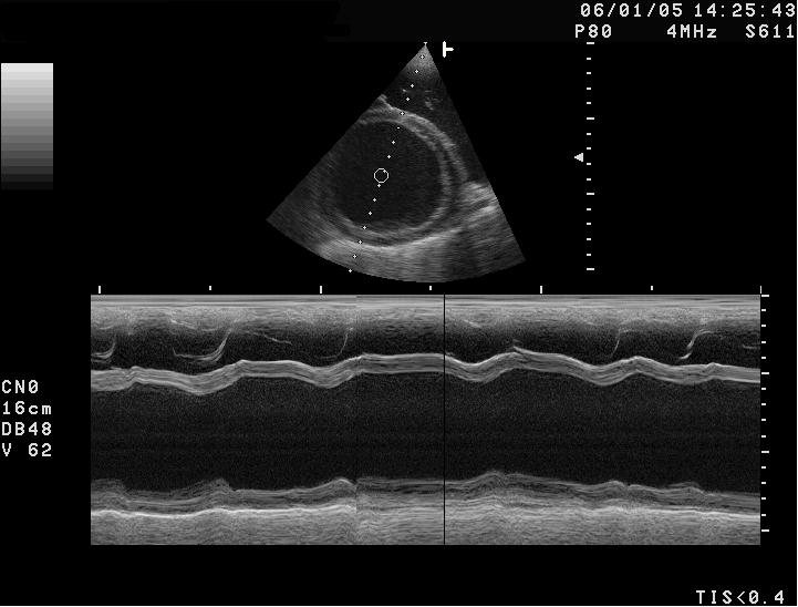
7-year-old Dobermann with exercise intolerance and premature beats on auscultation. Left ventricular M-mode echocardiogram (short axis); the calculated fractional shortening is 12%.
 zoom_inEnlarge
What is the most likely diagnosis?
chevron_right Show answer and explanation
A. Myxomatous mitral valve disease
In MMVD the ventricle can dilate, but systolic function stays normal or hyperdynamic (high fractional shortening), at least until very advanced stages.
B. Hypertrophic cardiomyopathy
HCM produces thickened walls and a small cavity, the opposite of the image. It is also very rare in dogs.
C. Dilated cardiomyopathy
Correct. A dilated left ventricle with hypokinetic walls and markedly reduced fractional shortening, in a Dobermann with ventricular arrhythmias: this is the dilated cardiomyopathy phenotype. In the occult phase, pimobendan (PROTECT study) delays heart failure and sudden death.
D. Pericardial effusion
Effusion would appear as an anechoic space around the heart, with rather small chambers due to compression.
lightbulb Key takeaway
In Dobermanns, screen yearly from 3 years of age with Holter (more than 300 ventricular premature complexes in 24 h is abnormal) and echocardiography. In atypical breeds (Golden Retrievers, dogs on legume-rich grain-free diets) consider diet-associated or taurine-deficiency cardiomyopathy, which may improve with a diet change.
Test yourself with 10 random questions
Cardiology · 100 questions, a different test every time
Related questions
Updated: October 31, 2026 · Educational content for veterinary professionals. It does not replace current clinical guidelines or the judgement of the attending veterinarian.