Dermatology
Random test · 10 questions
Question 1/10
7-year-old crossbreed dog, with no previous skin problems, with recent-onset pruritus and erythema. The lesions are confined to the ventral abdomen, scrotum, plantar pads and interdigital spaces, with a sharp border against haired skin. The floor cleaning product was changed 10 days ago.
What is the most likely diagnosis and how is it confirmed?
chevron_right Show answer and explanation
A. Atopic dermatitis; confirmed with allergen serology
Atopy usually starts between 6 months and 3 years of age, not suddenly at 7 years, and serology does not confirm the diagnosis.
B. Contact dermatitis (irritant or allergic); confirmed by avoiding the suspected product for 2–3 weeks and, if desired, re-exposing
Correct. Lesions confined to glabrous skin in contact with the floor, with a sharp border and a temporal link to a new product, are typical. Improvement on avoidance and relapse on re-exposure confirm the diagnosis; patch testing is an option in doubtful cases.
C. Sarcoptic mange; confirmed by superficial skin scraping
Mange typically affects the ear margins, elbows and hocks with intense pruritus and does not spare haired skin. Moreover, scrapings are frequently negative.
D. Superficial pyoderma; confirmed by bacterial culture
Pyoderma may complicate the picture, but it is secondary; the pattern confined to contact areas points to the primary cause.
lightbulb Key takeaway
Haired skin acts as a barrier, which is why contact dermatitis spares hairy areas and affects glabrous ones (abdomen, scrotum, pads, chin). Always ask about new products: cleaners, carpets, plastic bowls or topical treatments.
Question 2/10
6-year-old domestic shorthair cat on cefalexin for 8 days for a wound. It has fever, anorexia and painful erythema progressing to vesicles and epidermal sloughing on the trunk and limbs (over 50% of the surface), with oral ulcers. Rubbing apparently normal skin detaches the epidermis.
What is the most likely diagnosis and the correct approach?
chevron_right Show answer and explanation
A. Pemphigus vulgaris: start prednisolone and chlorambucil at immunosuppressive doses
Pemphigus vulgaris also causes mucocutaneous erosions, but its course is more gradual and it is not linked to a newly introduced drug. Histology distinguishes them (suprabasal acantholysis versus full-thickness epidermal necrosis).
B. Toxic epidermal necrolysis: withdraw the suspected drug and treat like a major burn
Correct. More than 30% of the surface involved with full-thickness epidermal necrosis, positive Nikolsky sign, mucosal involvement and fever. Withdraw all non-essential drugs and give intensive support: fluids, analgesia, nutrition and wound care. The prognosis is guarded to poor.
C. Erythema multiforme minor: it will resolve on its own without further measures
Erythema multiforme presents with target or annular lesions, affects less than 10% of the surface and has minimal mucosal involvement. Extensive sloughing with systemic signs belongs to the severe end of the spectrum.
D. Disseminated superficial pyoderma: culture and an antibiotic from another class
Pyoderma does not cause sheet-like epidermal sloughing or a Nikolsky sign. Moreover, the cat was already receiving an antibiotic, which is the main suspect.
lightbulb Key takeaway
Classification by extent of sloughing: Stevens-Johnson syndrome < 10%, overlap 10–30%, toxic epidermal necrolysis > 30%. The drugs most often involved in small animals are antibiotics (beta-lactams, potentiated sulphonamides). Record the reaction and do not give that drug or structurally related ones again.
Question 3/10
4-month-old Shetland Sheepdog with alopecia, erythema, scaling and crusts around the eyes, on the muzzle, ear margins, tail tip and over the carpi and tarsi. There is mild atrophy of the temporal and masseter muscles.
What is the most likely diagnosis?
chevron_right Show answer and explanation
A. Juvenile demodicosis
It is common at this age and should be ruled out with deep skin scrapings or trichograms, but it does not explain the lesions on the tail tip and bony prominences or the muscle atrophy.
B. Dermatophytosis
It can cause facial alopecia in puppies, but it does not produce scarring at pressure points or myositis. A fungal culture rules it out.
C. Discoid lupus erythematosus
It mainly affects the nasal planum of adults and is not accompanied by muscle atrophy or lesions on the tail tip and bony prominences of a puppy.
D. Familial dermatomyositis
Correct. It is a hereditary ischaemic dermatopathy of Collies, Shetland Sheepdogs and their crosses, beginning before 6 months of age on the face, ears, tail and friction areas, with variable myositis (masticatory muscles, sometimes megaoesophagus). It is confirmed by skin biopsy ± electromyography or muscle biopsy.
lightbulb Key takeaway
Treatment relies on pentoxifylline, avoiding sun and trauma, and vitamin E; severe cases may need immunomodulators. Lesions often leave alopecic scars. Advise against breeding affected dogs or their parents.
Question 4/10
11-year-old white cat with access to a sunny terrace. For months it has had erythema, scaling and crusts on the tips of both ears, which now bleed and have non-healing erosions.
Which statement is correct?
chevron_right Show answer and explanation
A. It is induced by ultraviolet radiation in poorly pigmented skin; it is locally invasive and pinnectomy with margins is often curative
Correct. It usually follows a phase of solar dermatitis and actinic keratosis in white, sparsely haired areas (ear tips, nasal planum, eyelids). It metastasises rarely and late, so local control is key: surgery, and on the nasal planum also cryotherapy, radiotherapy or electrochemotherapy depending on size.
B. It metastasises early to the lungs, so the prognosis is poor even after excision
Sun-induced feline cutaneous squamous cell carcinoma is mainly locally invasive. When detected early, surgery with adequate margins offers a good prognosis.
C. To prevent it, apply human sunscreens containing zinc oxide to the ears
The cat grooms and may ingest the product; zinc oxide and other ingredients can be toxic. Use sunscreens formulated for animals and, above all, avoid sun exposure in the middle of the day.
D. Crusts on the ear tips of a white cat are usually dermatophytosis: treat for 6 weeks before biopsying
Delaying biopsy allows the tumour to progress. Non-healing crusting and erosive lesions in white, sun-exposed areas should be biopsied early.
lightbulb Key takeaway
In Spain, with many hours of sunshine, white cats or cats with depigmented ears and noses that sunbathe are at high risk. Check their ears, nose and eyelids at every visit and biopsy any non-healing crusting lesion early: early lesions are cured with much less aggressive surgery.
Question 5/10
5-year-old Siamese cat with symmetrical alopecia on the ventral abdomen and caudal thighs, without erythema or crusts. The owners insist she does not scratch and suspect a hormonal problem.
On trichogram, the hairs have fractured tips and normal anagen and telogen bulbs. What is the correct interpretation?
chevron_right Show answer and explanation
A. Endocrine alopecia due to hyperadrenocorticism, typical of young cats
Hyperadrenocorticism is rare in cats, affects older animals, is usually associated with diabetes and fragile skin, and does not produce fractured tips.
B. Self-induced alopecia from excessive licking, almost always secondary to pruritus (allergy, fleas), even if the cat does it unseen
Correct. Broken tips indicate that the hair has been pulled or cut by the tongue. Many cats groom in secret. The cause of pruritus must be sought before considering a behavioural origin.
C. Psychogenic alopecia; clomipramine should be started
Psychogenic alopecia is rare and is only diagnosed after ruling out causes of pruritus, especially allergies. Treating with psychoactive drugs without a work-up leaves the real cause undiagnosed.
D. Telogen effluvium after a stressful episode
In telogen effluvium hair falls out whole, with most bulbs in telogen and intact tips, and the alopecia is usually more diffuse.
lightbulb Key takeaway
The trichogram is a quick, cheap test that distinguishes licking-induced alopecia (broken tips) from spontaneous hair loss. With self-induced alopecia, the work-up is that of the pruritic cat: flea control, elimination diet and then atopy.
Question 6/10
3-year-old Akita with progressive depigmentation of the nasal planum, lips and eyelids and some periocular leucotrichia. The owners have noticed red eyes, squinting and photophobia over the last few days.
What is the priority in this patient?
chevron_right Show answer and explanation
A. Urgent ophthalmic examination and systemic plus topical ocular immunosuppression without delay
Correct. This is uveodermatological (Vogt-Koyanagi-Harada-like) syndrome: an immune attack on the melanocytes of the skin and uvea. Panuveitis can cause blindness through synechiae, secondary glaucoma, cataracts or retinal detachment, so it is treated aggressively from day one.
B. Take skin biopsies and wait for the result before starting any treatment
Biopsy (lichenoid interface dermatitis with melanin-laden macrophages) supports the diagnosis, but can be taken while treatment starts. Waiting one or two weeks puts vision at risk.
C. Vitiligo: a cosmetic problem that needs no treatment
Vitiligo depigments without skin or ocular inflammation. Red eyes with facial depigmentation in an Akita should suggest uveodermatological syndrome.
D. Discoid lupus: topical tacrolimus on the nasal planum
It treats only the skin and overlooks the uveitis, which determines the prognosis. The skin lesions are the least serious part of this syndrome.
lightbulb Key takeaway
In Akitas, Siberian Huskies, Samoyeds and Chow Chows with facial depigmentation, always examine the eyes. Ocular signs often precede skin signs, and treatment is usually lifelong (systemic corticosteroids with azathioprine or other immunosuppressants, plus topical ocular therapy) with frequent ophthalmic monitoring.
Question 7/10
5-year-old Dachshund with several soft subcutaneous nodules on the trunk, 1 to 3 cm in size, some fistulated and draining a yellowish, oily fluid. It has mild fever and lethargy.
What is the most appropriate next diagnostic step?
chevron_right Show answer and explanation
A. 4 mm punch biopsy of the surface of a nodule
A small punch barely reaches the subcutaneous fat, where the lesion lies; the sample is often non-diagnostic.
B. Cytology of the exudate: if no organisms are seen, start prednisolone
Cytology shows pyogranulomatous inflammation, but failing to see organisms does not rule out mycobacteria, fungi or Nocardia, which may be scarce. Immunosuppressing such an infection would worsen it.
C. Excisional biopsy of an intact nodule for histopathology and tissue culture (bacteria, mycobacteria and fungi)
Correct. Sterile panniculitis is a diagnosis of exclusion: it requires a deep sample including fat, with special stains and negative tissue cultures. Choose a non-fistulated nodule to avoid contamination.
D. Empirical antibiotic for 6 weeks and assess the response
It delays diagnosis and does not cover mycobacteria or fungi. If the lesions are sterile, they will not respond.
lightbulb Key takeaway
Before diagnosing idiopathic sterile panniculitis rule out infections, foreign bodies, injection reactions, trauma and panniculitis associated with pancreatitis or pancreatic neoplasia. Idiopathic forms usually respond well to prednisolone; a single nodule may be cured by excision.
Question 8/10
6-year-old indoor domestic shorthair with feline atopic skin syndrome diagnosed by exclusion (flea control and elimination diet without response). It has received several depot corticosteroid injections in the past year.
Which option is most appropriate for long-term control?
chevron_right Show answer and explanation
A. Oral ciclosporin licensed for cats, avoiding hunting and raw meat because of the toxoplasmosis risk
Correct. Ciclosporin oral solution is licensed for chronic allergic dermatitis in cats. The main specific risk is primary Toxoplasma infection, so the cat should not hunt or eat raw meat.
B. Continue depot methylprednisolone acetate every 4–6 weeks
Repeated depots are associated with diabetes mellitus and congestive heart failure in cats. If glucocorticoids are used, oral (prednisolone, not prednisone) at the lowest effective dose is preferable.
C. Monthly lokivetmab, as in dogs
Lokivetmab is an antibody against canine IL-31, licensed only in dogs. It must not be extrapolated to cats.
D. Antihistamines as the sole treatment
Some cats respond partially, but their efficacy is limited and inconsistent. They can be adjuncts, not the basis of long-term control.
lightbulb Key takeaway
In cats, prefer prednisolone to prednisone (poorer hepatic conversion) and avoid depots. With ciclosporin, run baseline blood work, check FeLV/FIV status and reduce the frequency once controlled. Oclacitinib is not licensed in cats.
Question 9/10
14-year-old crossbreed with severe osteoarthritis and urinary incontinence that lives in a yard. In July the owner notices a bad smell: the perianal and perineal region has wounds with undermined edges full of moving white maggots. The dog is dull and somewhat dehydrated.
What is the most appropriate approach?
chevron_right Show answer and explanation
A. Apply an occlusive antibiotic ointment to suffocate the maggots and recheck in a week
Maggots continue to destroy tissue while alive and can migrate under the skin. Leaving them for a week worsens the lesion and the dog's condition; they must be removed now.
B. Sedation and analgesia, wide clipping, mechanical removal of all maggots, lavage and debridement, fluid therapy and correction of the incontinence and hygiene
Correct. The mainstay is removing all maggots (checking tracts under the skin), cleaning and debriding, and treating the patient (dehydration, pain, possible infection or toxaemia). Without addressing the urine and faeces that attract flies, it will recur.
C. Apply permethrin directly to the wounds to kill the maggots
Applying insecticides to open wounds promotes systemic absorption and tissue irritation. Dead maggots left in deep tracts also remain a source of infection.
D. Remove the maggots and close the wounds by primary suture in the same procedure
These are contaminated wounds with devitalised tissue and tracts. Immediate primary closure traps infection and possible residual maggots; they are managed open until a healthy granulation bed is present.
lightbulb Key takeaway
Myiasis is a summer emergency in animals with wounds, coats soiled with urine or faeces, incontinence or reduced mobility. Check at-risk patients daily during fly season and explain to the owner that hygiene is the best prevention.
Question 10/10
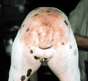
5-year-old dog with intense pruritus for 3 weeks, in summer. The owner has not seen any fleas. The lesions are shown in the image.
 zoom_inEnlarge
What is the most likely diagnosis?
chevron_right Show answer and explanation
A. Atopic dermatitis
Atopic dermatitis typically affects the face, ears, distal limbs, axillae, groin and ventrum, not predominantly the lumbosacral area.
B. Flea allergy dermatitis (FAD)
Correct. Lumbosacral, tail-base and caudal thigh distribution is the classic FAD pattern. The owner not seeing fleas does not rule it out: the allergic dog removes them by licking and scratching.
C. Sarcoptic mange
Sarcoptic mange mainly affects the ear margins, elbows, hocks and ventrum.
D. Dermatophytosis
In dogs, dermatophytosis usually causes focal, scaly alopecic lesions with variable pruritus, not such an intensely pruritic lumbosacral pattern.
lightbulb Key takeaway
Look for flea dirt with a comb and damp paper (it turns red). Treat every dog and cat in the household year-round with a fast-acting ectoparasiticide (isoxazolines) and treat the environment: most of the flea population lives in the house, not on the animal.
Review: open any question to study it
- Contact dermatitis in dogs: glabrous areas and avoidance trial
- Toxic epidermal necrolysis in cats: drug reaction
- Familial dermatomyositis in Collies and Shelties: diagnosis
- Squamous cell carcinoma in white cats: ears and nose
- Symmetrical alopecia in the cat: the trichogram and hidden licking
- Uveodermatological syndrome in Akitas: an ophthalmic emergency
- Sterile nodular panniculitis in dogs: how to diagnose it
- Treating the atopic cat: ciclosporin and corticosteroids
- Myiasis in dogs: treating maggot-infested wounds
- Flea allergy dermatitis (FAD) in dogs