Traumatic diaphragmatic hernia: radiographic findings
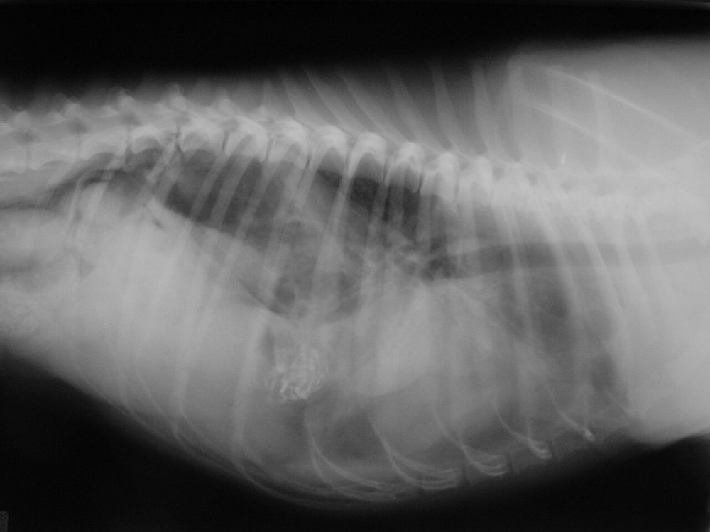
Dog hit by a car 3 days ago, presenting with progressive dyspnoea, exercise intolerance and an “empty” abdomen on palpation. Lateral thoracic radiograph:
 zoom_inEnlarge
What is the most likely diagnosis?
chevron_right Show answer and explanation
A. Traumatic pneumothorax
In pneumothorax the lungs retract from the thoracic wall and the heart lifts off the sternum, with free gas lacking vascular markings. It does not explain viscera inside the thorax or the empty abdomen.
B. Pulmonary contusion
Contusion produces a patchy alveolar or interstitial pattern that appears within hours and improves in 3–7 days. It does not erase the diaphragmatic line or bring gas-filled loops into the thorax.
C. Traumatic diaphragmatic hernia
Correct. The diaphragmatic outline is lost and gas- and content-filled structures (stomach, intestine) appear in the thorax, displacing heart and lungs. The “empty” abdomen on palpation supports the diagnosis.
D. Pleural effusion
Effusion produces interlobar fissure lines, scalloped lung margins and homogeneous fluid opacity obscuring the cardiac silhouette, without gas. (It can accompany a diaphragmatic hernia.)
lightbulb Key takeaway
Stabilise before surgery (oxygen, analgesia, sternal recumbency). If the stomach is herniated and dilating, it is an immediate surgical emergency. If in doubt, ultrasound or positive contrast confirms the diagnosis.
Test yourself with 10 random questions
Diagnostic imaging · 200 questions, a different test every time
Related questions
Updated: October 31, 2026 · Educational content for veterinary professionals. It does not replace current clinical guidelines or the judgement of the attending veterinarian.