Ascites in a young dog: hepatic arteriovenous malformation on CT
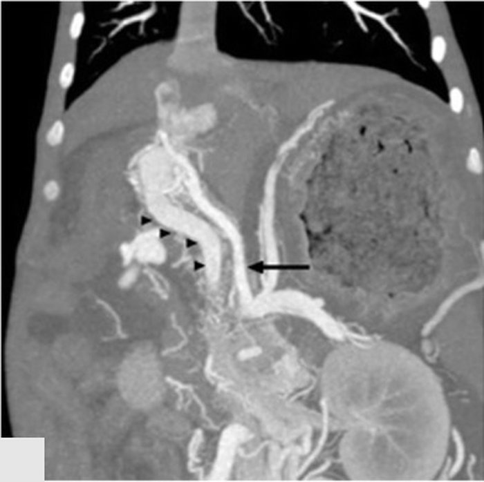
5-month-old male Labrador with weight loss and ascites. Bile acids are raised. On CT angiography, a markedly dilated artery arises from the coeliac trunk and the portal vein is already opacified, dilated and tortuous in the arterial phase. Dorsal reconstruction:
 zoom_inEnlarge
Which diagnosis explains these findings and the ascites?
chevron_right Show answer and explanation
A. Single large congenital extrahepatic portosystemic shunt
In a congenital shunt, portal blood escapes into the systemic circulation: the portal vein is usually small and ascites is rare. Here the portal vein is dilated and fills from a high-pressure artery.
B. Primary portal vein hypoplasia (microvascular dysplasia)
It features a small portal vein with no abnormal arterial communication. It can cause portal hypertension, but it does not explain a huge artery with early portal opacification.
C. Portal vein thrombosis secondary to a hypercoagulable state
Thrombosis causes a filling defect in a poorly opacified portal vein, not a large portal vein that fills in the arterial phase.
D. Hepatic arteriovenous malformation with portal hypertension
Correct. A congenital arterioportal communication delivers high-pressure arterial blood into the portal vein, which opacifies in the arterial phase, dilates and becomes arterialised. Portal hypertension follows, with ascites and multiple acquired shunts. It is mainly diagnosed in young dogs, and treatment is embolisation or lobectomy.
lightbulb Key takeaway
Ascites in a young dog with raised bile acids: think of portal hypertension (hepatic arteriovenous malformation, portal hypoplasia), not only a congenital shunt, which rarely causes ascites. CT angiography defines the type of vascular anomaly.
Test yourself with 10 random questions
Diagnostic imaging · 200 questions, a different test every time
Related questions
Updated: November 1, 2026 · Educational content for veterinary professionals. It does not replace current clinical guidelines or the judgement of the attending veterinarian.