Triceps tendon rupture in the dog: MRI findings
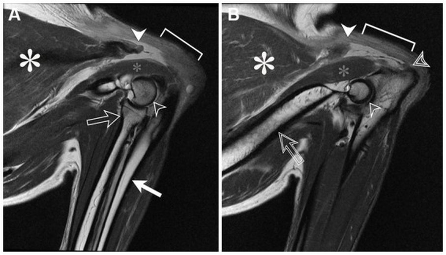
Active adult dog with acute right forelimb lameness; weeks later the same problem appears on the left. On weight bearing the elbow flexes excessively and the dog cannot keep it extended. Elbow radiographs are normal. Sagittal proton-density MRI of both elbows in flexion:
 zoom_inEnlarge
What is the diagnosis?
chevron_right Show answer and explanation
A. Bicipital tenosynovitis with effusion in the tendon sheath
The biceps tendon lies on the cranial aspect of the shoulder, not the caudal aspect of the elbow. Moreover, bicipital tendinopathy does not prevent the dog from keeping the elbow extended.
B. Triceps tendon rupture with proximal retraction
Correct. The triceps muscle ends abruptly and, distal to it, the hypointense band of the insertional tendon is missing, retracted proximally; on one side a stump remains on the olecranon. It explains the inability to extend the elbow under load.
C. Anconeus tear with an intact triceps tendon
The anconeus looks normal in both elbows; what is missing is the insertional tendon of the triceps. An isolated anconeus lesion would not produce this extension deficit.
D. Displaced avulsion fracture of the olecranon
An olecranon avulsion would appear on radiographs as a displaced bone fragment, and these were normal. The MRI shows an absent tendon, not a bone fragment.
lightbulb Key takeaway
Healthy tendons are hypointense on all MRI sequences. Their absence with a retracted stump, or increased T2 signal, indicates rupture. If the elbow drops on weight bearing and radiographs are normal, assess the triceps with ultrasound or MRI.
Test yourself with 10 random questions
Diagnostic imaging · 200 questions, a different test every time
Related questions
Updated: November 1, 2026 · Educational content for veterinary professionals. It does not replace current clinical guidelines or the judgement of the attending veterinarian.