Cruciate rupture in the cat: radiograph after surgery
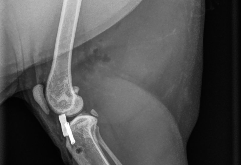
9-year-old overweight domestic shorthair cat operated on for a cranial cruciate ligament rupture in the right stifle. Mediolateral follow-up radiograph after surgery:
 zoom_inEnlarge
Which technique does this follow-up radiograph show?
chevron_right Show answer and explanation
A. TPLO: tibial plateau osteotomy fixed with a plate
There is no curved osteotomy of the proximal tibia and no plate with screws. TPLO is rarely used in cats.
B. TTA: tibial tuberosity advancement with cage and plate
In a TTA the tibial tuberosity is seen separated and advanced, with a cage in the osteotomy and a fork plate. Here the tuberosity is in its normal position.
C. Lateral fabellotibial suture with crimp and tibial tunnel
Correct. A metal crimp securing the suture and the tunnel drilled in the proximal tibia are visible. The nylon line is not radiopaque, so its path from the lateral fabella cannot be seen. It is the surgical technique most used in cats.
D. Lateral capsular imbrication with cerclage wire
Cerclage wire would appear as a continuous metal line around the joint. Here there is only a small compact implant next to a bone tunnel.
lightbulb Key takeaway
After a lateral fabellotibial suture, only the crimp and the tibial tunnel are visible on radiographs: suture integrity and stability are assessed on examination (drawer, tibial compression). In cats, also look for meniscal mineralisation, which is common in stifles with cruciate rupture.
Test yourself with 10 random questions
Diagnostic imaging · 200 questions, a different test every time
Related questions
Updated: November 1, 2026 · Educational content for veterinary professionals. It does not replace current clinical guidelines or the judgement of the attending veterinarian.