The five radiographic opacities: how to recognise fat
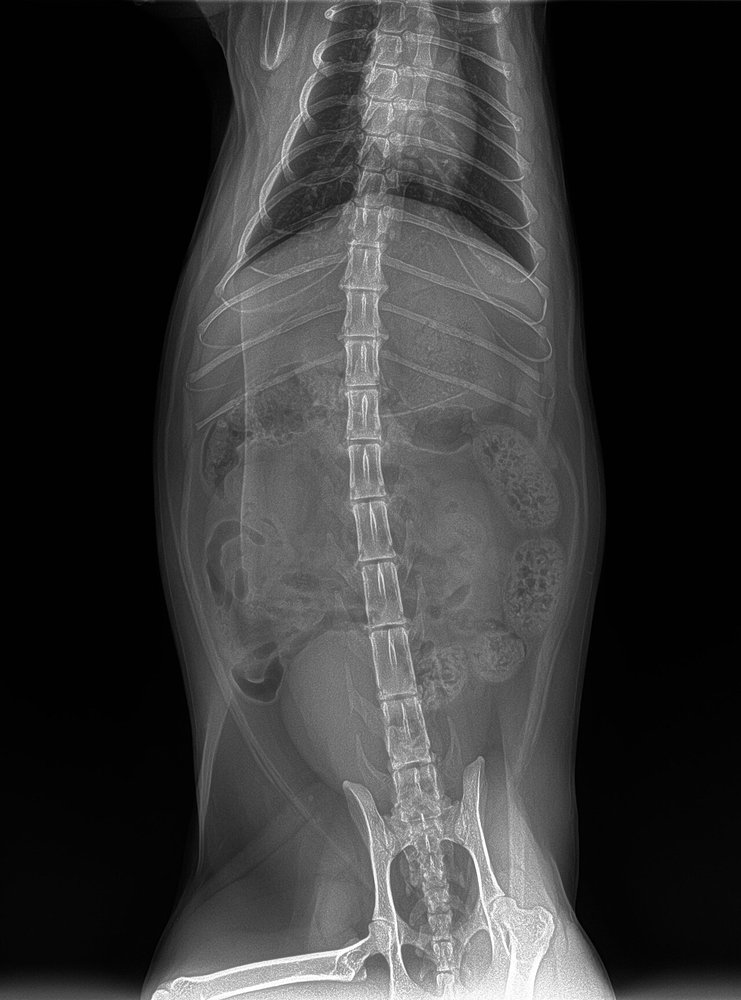
12-year-old neutered male cat, overweight (body condition 7/9), radiographed for suspected ingestion of a bone. Between the skin and the abdominal wall muscles there is a wide band, less opaque than muscle but more opaque than intestinal gas. Ventrodorsal radiograph:
 zoom_inEnlarge
What opacity does that band have, and what effect does abundant fat have on the image?
chevron_right Show answer and explanation
A. Gas opacity: subcutaneous emphysema that blurs margins
Gas is the most radiolucent opacity, almost as black as the surrounding air; emphysema produces bubbles or irregular streaks, not a homogeneous layer.
B. Soft tissue/fluid opacity: oedema that reduces contrast
Soft tissue and fluid have the same opacity as muscle and cannot be told apart from it; this band is less opaque than the muscles.
C. Mineral opacity: calcinosis that hides the viscera
Mineral is as opaque as bone or more so; the band is clearly less opaque than muscle.
D. Fat opacity: abdominal fat improves serosal contrast
Correct. Fat is the second most radiolucent opacity, between gas and soft tissue. In an overweight cat, intra-abdominal fat surrounds the viscera and outlines their margins; in very thin or young animals, serosal detail is poorer.
lightbulb Key takeaway
The five opacities, from least to most opaque: gas, fat, soft tissue/fluid, mineral/bone and metal. A margin is only seen where two different opacities meet: that is why fat outlines organs and peritoneal fluid obliterates them. A subcutaneous mass of fat opacity suggests a lipoma, but confirm it with cytology.
Test yourself with 10 random questions
Diagnostic imaging · 200 questions, a different test every time
Related questions
Updated: October 31, 2026 · Educational content for veterinary professionals. It does not replace current clinical guidelines or the judgement of the attending veterinarian.