Overexposure in digital radiography: burn-out and dose
4 kg cat. Lateral digital abdominal radiograph taken with the technique you use for medium-sized dogs. The exposure index is well above the manufacturer's range; the skin, tail and abdominal margins look completely black with no detail, although the rest of the image looks 'fine'. Repeat with technique adapted to thickness:
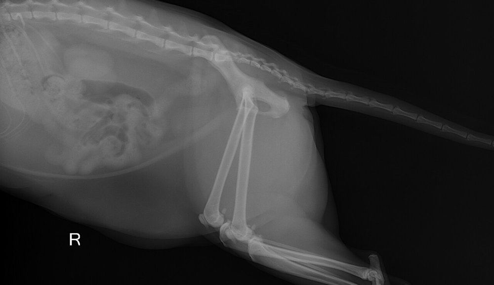 zoom_inEnlarge
What is the cause and what should you do?
chevron_right Show answer and explanation
A. Overexposure with detector saturation: repeat with lower mAs and kVp adapted to thickness
Correct. Digital processing rescales the image and hides overexposure, but in thin areas the detector saturates ('burn-out') and information is lost. Adjust technique to the measured thickness and monitor the exposure index.
B. Underexposure: mAs should be increased
Underexposure on digital systems gives a grainy image (quantum mottle) and a low exposure index, not saturated black areas with a high index.
C. No need to repeat: adjusting window and level recovers the lost detail
Saturated pixels contain no recoverable information; post-processing cannot bring it back. Accepting overexposure also encourages progressive dose increase ('dose creep').
D. Grid cut-off from an off-centre beam
Grid cut-off produces light, underexposed areas (usually at the sides), not black overexposed areas.
lightbulb Key takeaway
On digital systems a 'nice' image does not guarantee correct technique: always check the exposure index. Overexposure = unnecessary dose and loss of detail in thin areas; underexposure = noise. Use a technique chart based on measured thickness.
Test yourself with 10 random questions
Diagnostic imaging · 200 questions, a different test every time
Related questions
Updated: October 31, 2026 · Educational content for veterinary professionals. It does not replace current clinical guidelines or the judgement of the attending veterinarian.