Hip dysplasia: radiographs and the Norberg angle
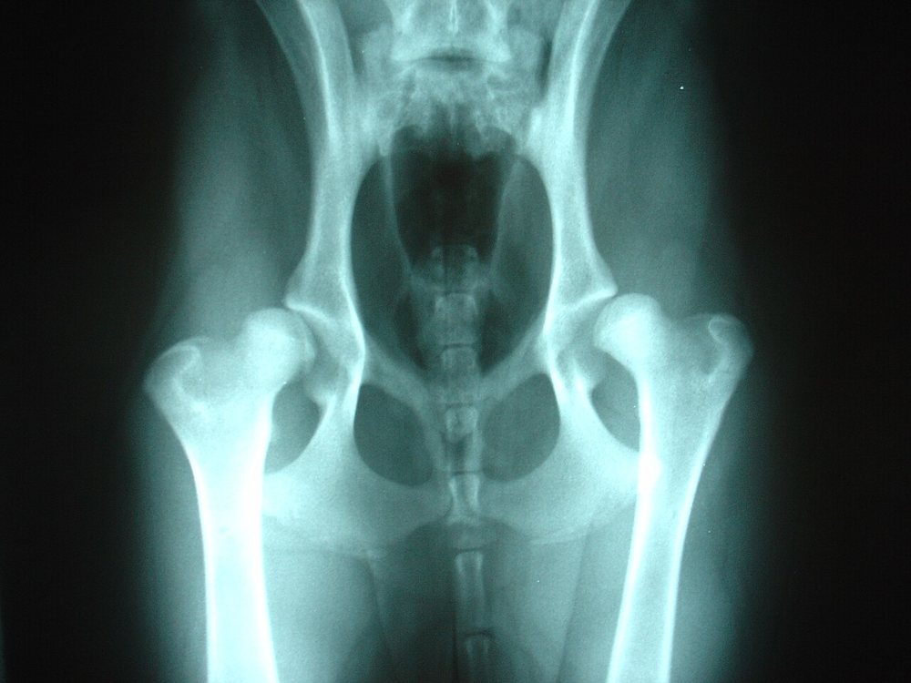
14-month-old Labrador with intermittent bilateral hindlimb lameness that worsens after exercise, and difficulty getting up. Ventrodorsal hip-extended pelvic radiograph:
 zoom_inEnlarge
Which statement about radiographic assessment of hip dysplasia is correct?
chevron_right Show answer and explanation
A. A Norberg angle greater than 115° indicates dysplasia
It is the other way round: the larger the angle, the better the femoral head is covered. An angle ≥ 105° is considered normal; low values indicate subluxation.
B. A lateral pelvic view is enough
The lateral view is useful for other lesions, but dysplasia assessment requires a ventrodorsal view with the limbs extended and parallel, patellae centred and the pelvis symmetrical, usually under sedation.
C. Signs of dysplasia: Norberg angle < 105°, subluxation with joint incongruity, a shallow acetabulum and osteophytes
Correct. In the image both femoral heads are poorly covered by the acetabula (bilateral subluxation). Over time femoral neck remodelling, the Morgan line and osteophytes appear due to degenerative joint disease.
D. Dysplasia cannot be assessed radiographically before 5 years of age
Official FCI assessment is done from 12 months (18 in giant breeds), and the PennHIP joint laxity method can be used from 16 weeks of age.
lightbulb Key takeaway
Radiographic severity does not always match the clinical signs: treat the patient, not the radiograph. Options: weight control, controlled exercise, NSAIDs or anti-NGF antibodies, physiotherapy and, in selected cases, surgery (juvenile pubic symphysiodesis, TPO or hip replacement).
Test yourself with 10 random questions
Diagnostic imaging · 200 questions, a different test every time
Related questions
Updated: October 31, 2026 · Educational content for veterinary professionals. It does not replace current clinical guidelines or the judgement of the attending veterinarian.