Bladder stones on ultrasound: acoustic shadowing and diagnosis
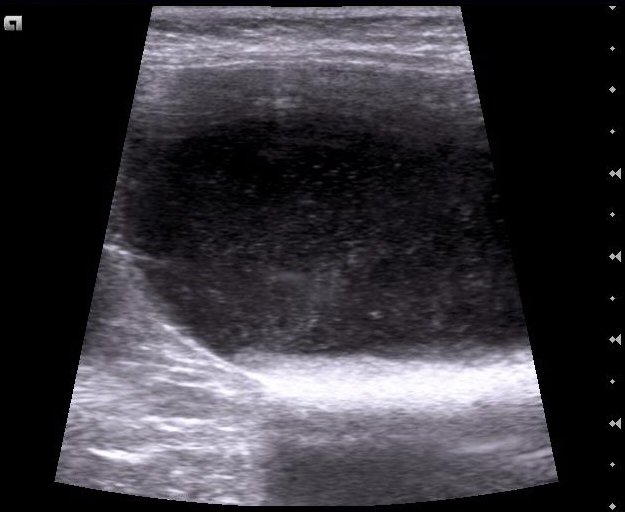
1-year-old male cat with urethral obstruction. On ultrasound the bladder is distended and contains fine suspended echoes; along the dependent wall there is a hyperechoic layer that moves when the patient is repositioned and casts an acoustic shadow:
 zoom_inEnlarge
What is that hyperechoic layer most likely to be?
chevron_right Show answer and explanation
A. Bladder wall thickening or polyp
A polyp is attached to the wall, does not move with gravity, casts no acoustic shadow and usually shows blood flow on colour Doppler.
B. Mineral concretions: sand-like sediment or small uroliths
Correct. A hyperechoic surface, dependent position, movement with changes of position and a clean acoustic shadow are the ultrasound criteria for mineral material. In male cats, sand-like sediment and small uroliths can obstruct the urethra.
C. Blood clot
A clot may move, but it is usually heterogeneous, of medium echogenicity, and casts no acoustic shadow.
D. Cellular or proteinaceous sediment without a mineral component
Cellular or proteinaceous sediment settles in the dependent area and resuspends when the bladder is balloted, but it is less echogenic and does not produce a clean acoustic shadow.
lightbulb Key takeaway
Clean acoustic shadow + movement with gravity = mineral material. The colour Doppler twinkling artefact helps with small stones. Add radiographs of the whole urinary tract, urethra included: urate and cystine uroliths may be radiolucent and seen only on ultrasound.
Test yourself with 10 random questions
Diagnostic imaging · 200 questions, a different test every time
Related questions
Updated: October 31, 2026 · Educational content for veterinary professionals. It does not replace current clinical guidelines or the judgement of the attending veterinarian.