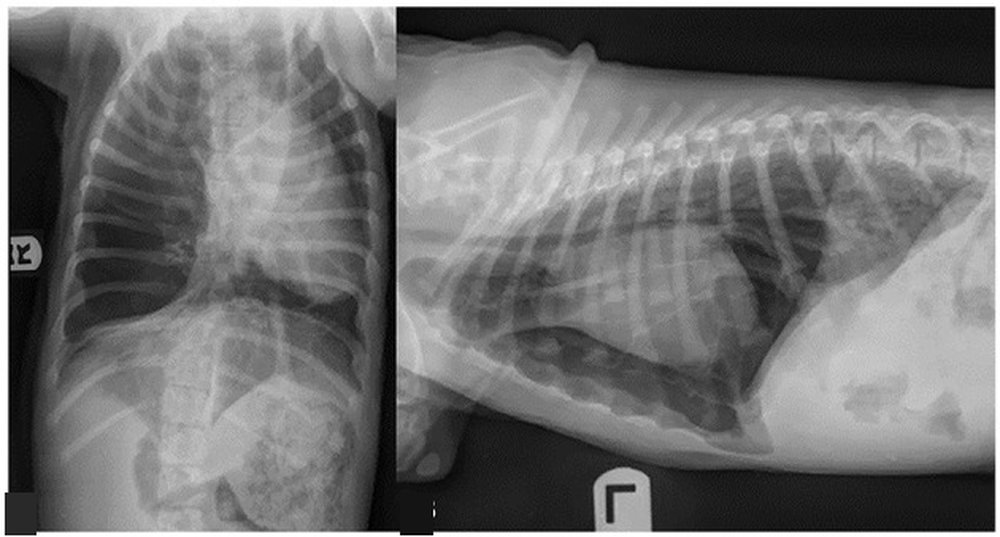
Congenital lobar emphysema in the puppy: radiograph
11-week-old female Catahoula puppy with progressive tachypnoea and respiratory distress over the past week, with no history of trauma. Ventrodorsal and lateral thoracic radiographs:
 zoom_inEnlarge
A right lung lobe is markedly hyperlucent and enlarged, with the mediastinum and heart shifted to the left. What is the most likely diagnosis?
chevron_right Show answer and explanation
A. Congenital lobar emphysema
Correct. An overinflated lobe in a young animal, with contralateral mediastinal shift and vascular markings still visible within the hyperlucent area, is typical of congenital lobar emphysema. CT confirms it and lobectomy is curative.
B. Right tension pneumothorax
In pneumothorax the lucent area lacks vascular markings and the collapsed lung retracts from the wall. Here the lobe still contains vessels and the course is progressive rather than acute.
C. Congenital diaphragmatic hernia
A gas-filled viscus in the thorax would show a gastric wall or bowel loops and loss of the diaphragmatic line. Here the diaphragm is intact.
D. Giant congenital pulmonary bulla
A bulla is a thin-walled gas cavity with no vessels inside, usually rounded. It does not typically overinflate a whole lobe while keeping its vascular pattern.
lightbulb Key takeaway
With a hyperlucent hemithorax, look for vessels within the dark area: if present, it is overinflated lung rather than free gas. In a puppy with progressive dyspnoea, think of congenital lobar emphysema and do not place a chest drain before confirming it with CT.
Test yourself with 10 random questions
Diagnostic imaging · 200 questions, a different test every time
Related questions
Updated: November 1, 2026 · Educational content for veterinary professionals. It does not replace current clinical guidelines or the judgement of the attending veterinarian.