Medial patellar luxation in dogs: what radiographs add
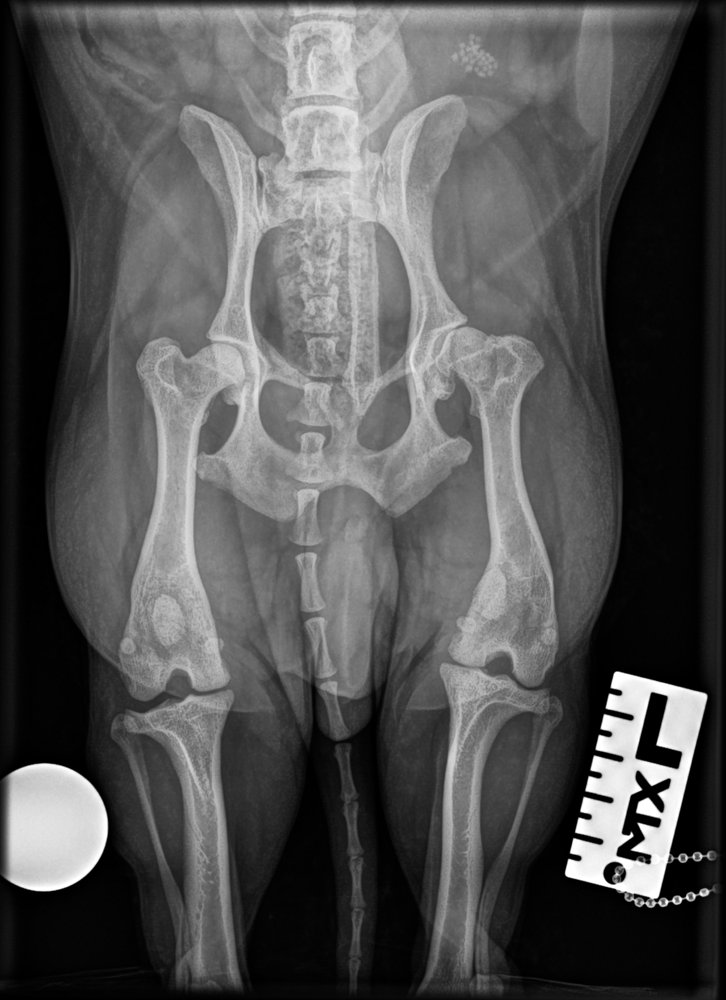
2-year-old dog with intermittent pelvic limb lameness and 'skipping', carrying a limb for a few strides. On palpation both patellae luxate medially with ease and one stays luxated most of the time. Ventrodorsal radiograph of the pelvis and pelvic limbs:
 zoom_inEnlarge
Which statement about the radiographic work-up is correct?
chevron_right Show answer and explanation
A. It assesses bone deformities, trochlear depth and osteoarthritis; the grade is set by palpation
Correct. Grading I–IV is clinical. Radiographs (and CT if needed) show distal femoral varus, tibial torsion, tuberosity position and osteoarthritis, which determine the surgical technique.
B. Radiographs are essential to grade the patellar luxation
The grade depends on whether the patella luxates spontaneously or on manipulation and whether it reduces, which can only be assessed by palpation.
C. If the patella is in place on the radiograph, luxation is ruled out
In intermittent luxations (grades I–II) the patella is usually in the trochlear groove when the image is taken. A normal radiograph does not rule it out.
D. In small-breed dogs patellar luxation is almost always lateral
It is the other way round: medial luxation clearly predominates in small and toy breeds, often bilaterally. Lateral luxation is more typical of some large breeds.
lightbulb Key takeaway
Always examine both stifles: medial luxation is often bilateral. In high grades, assess femoral and tibial deformities on imaging, because the technique depends on them (trochleoplasty, tuberosity transposition or femoral osteotomy). Also look for effusion suggesting a concurrent cruciate rupture.
Test yourself with 10 random questions
Diagnostic imaging · 200 questions, a different test every time
Related questions
Updated: October 31, 2026 · Educational content for veterinary professionals. It does not replace current clinical guidelines or the judgement of the attending veterinarian.