Magnification and distortion in radiography: how to minimise them
Young dog lame on a pelvic limb after a fall. On this lateral radiograph the two pelvic limbs are not superimposed: the one further from the detector, tilted over the other, looks somewhat larger and less sharp than the one resting on the table. Lateral radiograph of the hips, femurs and stifles:
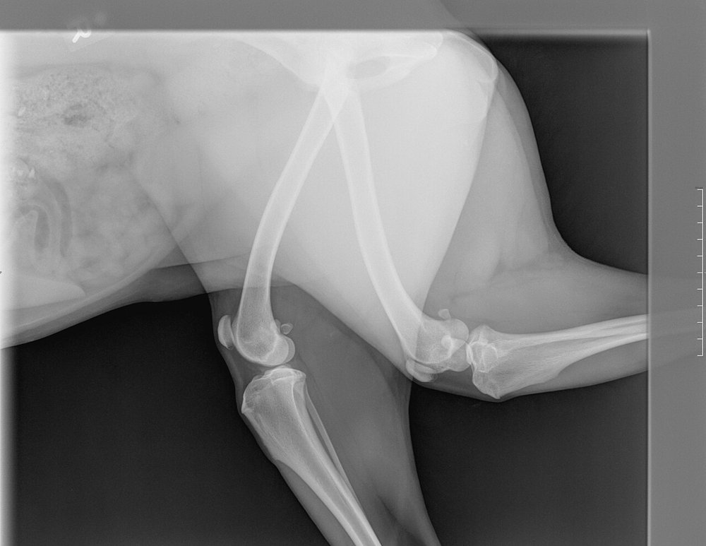 zoom_inEnlarge
Which measure best corrects these faults?
chevron_right Show answer and explanation
A. Shorten the focus–detector distance so the beam is more intense
A short focus–detector distance increases magnification and geometric unsharpness. To minimise them, use the greatest practical distance (usually ~100 cm).
B. Rest the limb directly on the detector, parallel to it, with the beam centred and perpendicular
Correct. The greater the object–detector distance, the greater the magnification and blurring. Distortion (change in shape) appears when the object is not parallel to the detector or the beam is oblique, because each part is magnified differently.
C. Increase mAs to improve edge sharpness
mAs alters exposure, not geometry. Blurring from object–detector distance or motion is not corrected by raising mAs.
D. Angle the tube so the beam follows the axis of the tilted bone
A beam oblique to the detector increases distortion (elongation or foreshortening). The correct approach is to place the bone parallel to the detector and the beam perpendicular.
lightbulb Key takeaway
For a faithful image: object close to and parallel with the detector, beam perpendicular and centred, a constant and sufficient focus–detector distance. That is why, in the thorax, the side nearest the detector appears sharper, and measurements (e.g. VHS) need a standardised technique.
Test yourself with 10 random questions
Diagnostic imaging · 200 questions, a different test every time
Related questions
Updated: October 31, 2026 · Educational content for veterinary professionals. It does not replace current clinical guidelines or the judgement of the attending veterinarian.