Aspiration pneumonia: alveolar pattern and air bronchograms
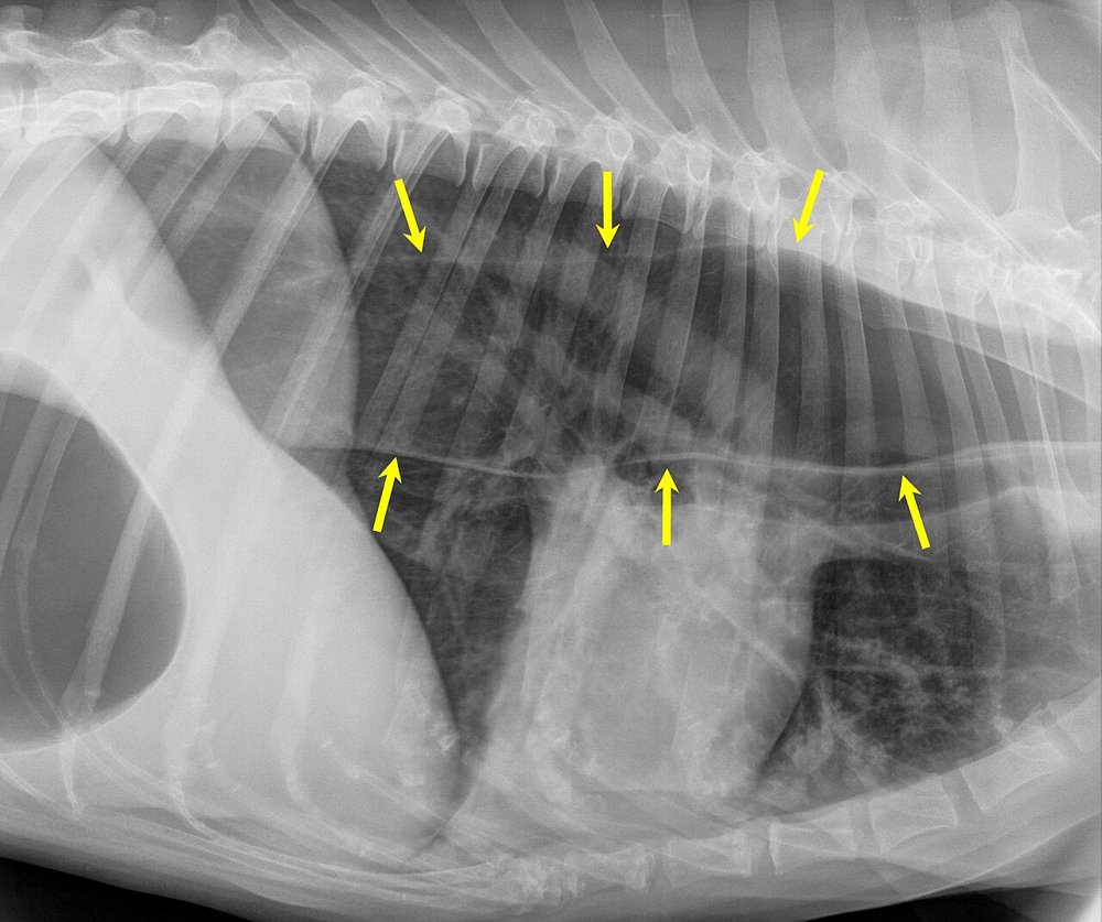
6-year-old dog with megaoesophagus (the yellow arrows mark its walls) and frequent regurgitation, now with a 2-day history of 39.9 °C fever, moist cough and tachypnoea. Lateral thoracic radiograph:
 zoom_inEnlarge
Apart from the megaoesophagus, which lung finding do you look for as a sign of aspiration pneumonia?
chevron_right Show answer and explanation
A. Diffuse bronchial pattern with pulmonary hyperinflation
This is the typical picture of lower airway disease (such as feline asthma or chronic bronchitis), not of pneumonia with alveolar filling.
B. Alveolar pattern with air bronchograms in the cranioventral lobes, especially the right middle lobe
Correct. Aspirated material goes to the dependent regions; the right middle lobe is the most affected because of the anatomy of the right principal bronchus. The air bronchogram (an air-filled bronchus surrounded by fluid-filled alveoli) defines the alveolar pattern.
C. Perihilar and caudodorsal alveolar pattern
That is the typical distribution of cardiogenic oedema in the dog, usually accompanied by cardiomegaly and left atrial enlargement.
D. Multiple well-defined interstitial nodules
A structured (nodular) interstitial pattern suggests metastases, granulomas or fungal disease, not aspiration pneumonia.
lightbulb Key takeaway
Cranioventral alveolar pattern = think bronchopneumonia or aspiration. Radiographic changes may lag behind clinical signs in the first hours. Always look for the cause of aspiration: megaoesophagus, laryngeal paralysis, brachycephalic syndrome or recent anaesthesia.
Test yourself with 10 random questions
Diagnostic imaging · 200 questions, a different test every time
Related questions
Updated: October 31, 2026 · Educational content for veterinary professionals. It does not replace current clinical guidelines or the judgement of the attending veterinarian.