Megaoesophagus in dogs: radiographs and complications
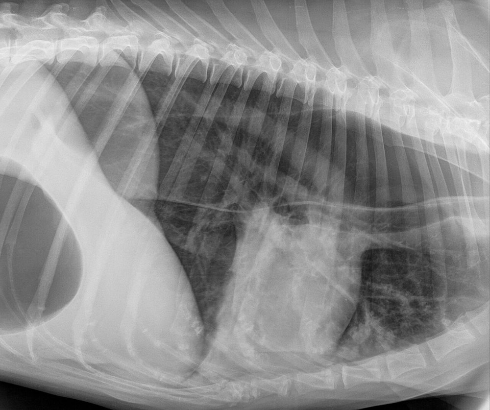
8-year-old dog that, according to the owner, “vomits” food without abdominal effort a few minutes after eating. It has lost weight. Lateral thoracic radiograph:
 zoom_inEnlarge
What is the main finding and its most common complication?
chevron_right Show answer and explanation
A. Pneumomediastinum; complication: pneumothorax
Pneumomediastinum outlines mediastinal structures (vessels, the outer tracheal wall) diffusely with gas, not as a dilated tube along the thorax. It would not explain the regurgitation either.
B. Hiatal hernia; complication: reflux oesophagitis
A hiatal hernia appears as a soft-tissue or gas opacity in the caudodorsal thorax next to the diaphragm, not as an oesophagus dilated along its whole length. It is more common in brachycephalic breeds.
C. Cardiogenic pulmonary oedema; complication: respiratory failure
There is no perihilar alveolar pattern or cardiomegaly. What stands out is a gas-filled tubular structure, and the history is regurgitation, not cough or dyspnoea.
D. Megaoesophagus; complication: aspiration pneumonia
Correct. The gas-dilated oesophagus is seen dorsal to the trachea, sometimes with the “tracheal stripe” sign. The history is regurgitation (passive, effortless), not vomiting. Aspiration pneumonia is the most common complication and the main cause of death.
lightbulb Key takeaway
In acquired adult megaoesophagus look for a cause: myasthenia gravis (acetylcholine receptor antibodies), hypoadrenocorticism, hypothyroidism or polymyopathies. Feed upright (Bailey chair) with balls of wet food.
Test yourself with 10 random questions
Diagnostic imaging · 200 questions, a different test every time
Related questions
Updated: October 31, 2026 · Educational content for veterinary professionals. It does not replace current clinical guidelines or the judgement of the attending veterinarian.