Cardiogenic pulmonary oedema on canine radiographs
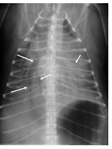
11-year-old Cavalier King Charles with a known mitral systolic murmur, now presenting with cough, resting tachypnoea (60 breaths/min) and respiratory effort. Dorsoventral radiograph; the arrows point to air bronchograms:
 zoom_inEnlarge
Which combination of radiographic findings is typical of cardiogenic pulmonary oedema in the dog?
chevron_right Show answer and explanation
A. Alveolar pattern in the cranioventral lobes
A cranioventral distribution (right middle and cranial lobes) is typical of aspiration pneumonia and bronchopneumonia, not cardiogenic oedema.
B. Diffuse bronchial pattern with “doughnuts” and “tram lines”
A bronchial pattern means thickened bronchial walls: chronic bronchitis, feline asthma. It can coexist with heart disease in small dogs, but it is not the sign of oedema.
C. Perihilar and caudodorsal interstitial–alveolar pattern, with left atrial enlargement and distended pulmonary veins
Correct. In dogs, cardiogenic oedema starts in the perihilar and caudodorsal region, with left-sided cardiomegaly and pulmonary venous congestion (veins wider than their paired arteries). In severe cases like this one it spreads and becomes alveolar, with air bronchograms.
D. Diffuse miliary nodular pattern
Diffuse miliary nodules suggest metastases, systemic mycoses or granulomatous pneumonia, not oedema.
lightbulb Key takeaway
In cats cardiogenic oedema is irregular and variable (often patchy, not perihilar) and pleural effusion is more common. Resting respiratory rate at home (> 30 breaths/min) is a great monitoring tool.
Test yourself with 10 random questions
Diagnostic imaging · 200 questions, a different test every time
Related questions
Updated: October 31, 2026 · Educational content for veterinary professionals. It does not replace current clinical guidelines or the judgement of the attending veterinarian.