Pulmonary osteomas on canine radiographs: not metastases
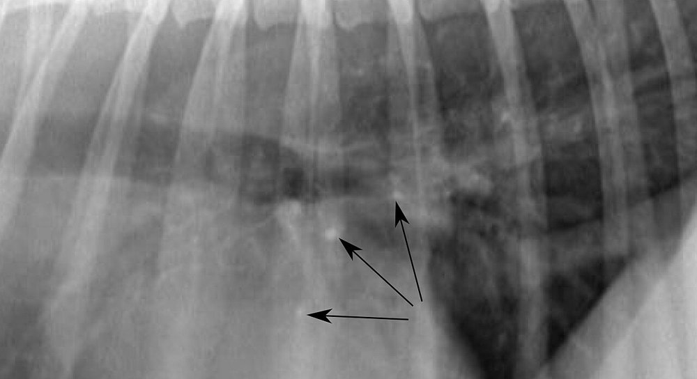
11-year-old dog being staged for a skin tumour. The lateral radiograph (detail) shows several pinpoint foci of 1–3 mm, with mineral opacity and very sharp margins (arrows):
 zoom_inEnlarge
What is the most likely interpretation?
chevron_right Show answer and explanation
A. Miliary metastases: the tumour has already spread
Metastatic nodules usually have soft-tissue opacity and need to be about 3–5 mm to be seen; foci this small and this opaque indicate mineral, not soft tissue.
B. Pulmonary osteomas, a common incidental finding in older dogs
Correct. They are small foci of heterotopic bone in the lung: very opaque for their size, with sharp and sometimes irregular margins, and more common in older and large-breed dogs. They are clinically insignificant.
C. Pulmonary vessels seen end-on, in cross-section
An end-on vessel also looks like an opaque dot, but it matches the calibre of its vessel and lies next to its bronchus; osteomas appear isolated and away from vascular tracts.
D. Mineralisation of the bronchial walls
Bronchial mineralisation, common in older dogs or those with hyperadrenocorticism, forms rings and parallel lines along the bronchi, not isolated pinpoint foci.
lightbulb Key takeaway
Mineral opacity, very small size and sharp margins in an older dog: think pulmonary osteoma. A typical metastatic nodule has soft-tissue opacity; if in doubt, compare with previous radiographs or use CT.
Test yourself with 10 random questions
Diagnostic imaging · 200 questions, a different test every time
Related questions
Updated: October 31, 2026 · Educational content for veterinary professionals. It does not replace current clinical guidelines or the judgement of the attending veterinarian.