Gastric dilatation-volvulus: which radiographic view to use
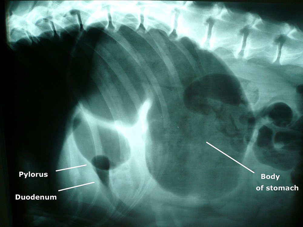
5-year-old Great Dane with abdominal distension, unproductive retching and hypersalivation. Already stabilised with fluids and gastric decompression. The image shows a lateral radiograph of a dog with a severely gas-distended stomach.
 zoom_inEnlarge
Which radiographic view is preferred to confirm gastric dilatation-volvulus (GDV)?
chevron_right Show answer and explanation
A. Right lateral (patient in right lateral recumbency)
Correct. In GDV the pylorus moves dorsally and to the left. With the patient on its right side, gas rises into the displaced pylorus, seen as a bubble separated from the fundus by a soft-tissue band: “compartmentalisation” or the “reverse C” sign.
B. Left lateral
In left recumbency the displaced pylorus is dependent and fills with fluid, so compartmentalisation is much less obvious. It is the best view for the pylorus in a normal stomach, not in GDV.
C. Ventrodorsal
Placing a GDV patient in dorsal recumbency worsens venous return and ventilation, and the view is less diagnostic than the right lateral.
D. Abdominal ultrasound instead of radiography
Gastric gas prevents assessing stomach position on ultrasound. Radiography is the test of choice.
lightbulb Key takeaway
Stabilise first, then radiograph. If the right lateral is inconclusive, add a left lateral or a dorsoventral view. Simple dilatation without volvulus shows no compartmentalisation.
Test yourself with 10 random questions
Diagnostic imaging · 200 questions, a different test every time
Related questions
Updated: October 31, 2026 · Educational content for veterinary professionals. It does not replace current clinical guidelines or the judgement of the attending veterinarian.