Pyometra in the bitch: radiographs and ultrasound confirmation
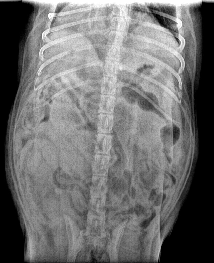
8-year-old intact bitch with lethargy, polyuria/polydipsia and anorexia, 6 weeks after her last season. No visible vulvar discharge. Ventrodorsal abdominal radiograph:
 zoom_inEnlarge
What is the most likely diagnosis and how would you confirm it?
chevron_right Show answer and explanation
A. Pregnancy of about 6 weeks: no further tests needed
A pregnant uterus also looks tubular, but the bitch would not be ill or PU/PD. Fetal skeletons do not mineralise until day 42–45; before that, radiographs cannot tell pregnancy from pyometra.
B. Closed pyometra: confirm with ultrasound
Correct. Soft-tissue tubular structures in the caudal and mid abdomen of a sick intact bitch in the luteal phase (dioestrus) suggest a distended uterus. Ultrasound confirms a uterus filled with anechoic or echogenic content and rules out pregnancy.
C. Mechanical intestinal obstruction
In obstruction you see small intestinal loops dilated with gas or fluid, often “stacked”, and there is usually vomiting. Here the structures are soft tissue without gas and the history points to the reproductive tract.
D. Ascites
Ascites produces a diffuse loss of serosal detail (“ground glass” appearance), not well-defined tubular structures.
lightbulb Key takeaway
Pyometra is an emergency. PU/PD occurs because E. coli endotoxins impair the response to ADH. The treatment of choice is ovariohysterectomy after stabilisation. Closed pyometra carries a higher risk of sepsis and uterine rupture than open pyometra.
Test yourself with 10 random questions
Diagnostic imaging · 200 questions, a different test every time
Related questions
Updated: October 31, 2026 · Educational content for veterinary professionals. It does not replace current clinical guidelines or the judgement of the attending veterinarian.