How to orient a ventrodorsal radiograph for reading
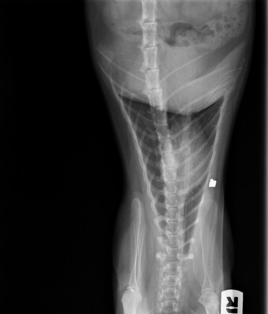
4-year-old Siamese cat with air-gun pellet wounds. When you open its ventrodorsal radiograph of the thorax and cranial abdomen in the viewer, it appears like this (the “R” marker shows the patient's right side):
 zoom_inEnlarge
What should you do to display it according to the standard convention?
chevron_right Show answer and explanation
A. Nothing: in VD views the caudal end is placed at the top
The convention is the opposite: in VD and DV views the cranial end goes at the top.
B. Rotate it 180°: cranial at the top and the patient's right on the viewer's left
Correct. VD and DV views are displayed with the cranial end at the top and the patient's right on the viewer's left, as if the animal were facing you. After a 180° rotation, the “R” marker sits at the top left and reads the right way round.
C. Flip it vertically so cranial is at the top without changing the side
With a vertical flip the patient's right would stay on the viewer's right, contrary to the convention, and the image would become a mirror image.
D. Flip it horizontally so the right side is on the viewer's left
It corrects the side but leaves the cranial end at the bottom, and flipping creates a mirror image that invites laterality errors.
lightbulb Key takeaway
Convention: on lateral views, the animal's head to the viewer's left; on VD and DV views, cranial at the top and the patient's right on the viewer's left. Always place a radiopaque marker at the time of exposure, not a digital annotation afterwards.
Test yourself with 10 random questions
Diagnostic imaging · 200 questions, a different test every time
Related questions
Updated: October 31, 2026 · Educational content for veterinary professionals. It does not replace current clinical guidelines or the judgement of the attending veterinarian.