Air-gun pellet over a cat's thorax: localising it on radiographs
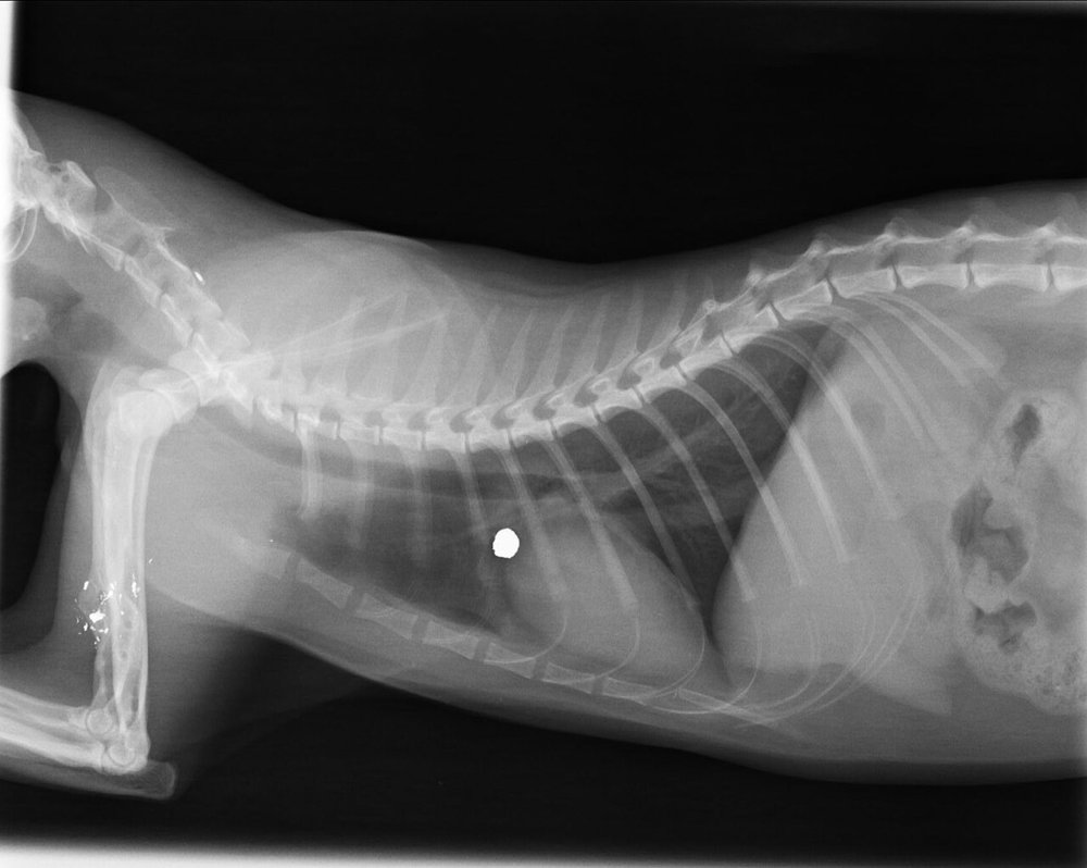
4-year-old Siamese cat found as a stray with air-gun pellet wounds; breathing is normal. There are metallic fragments in the elbow region and, on the lateral thoracic radiograph, a pellet is projected over the lung field:
 zoom_inEnlarge
What do you do to find out whether the pellet is inside the thorax?
chevron_right Show answer and explanation
A. An orthogonal view (VD or DV) to place it in the missing plane
Correct. A radiograph is a flat image: everything the beam passes through is superimposed. A view at 90° places the object in the third plane. In this cat, the ventrodorsal view showed the pellet in the soft tissues of the thoracic wall, not in the lung.
B. No further test: it is in the lung because it overlies it on the lateral
Overlying is not the same as lying within. On a single view, an object in the skin, thoracic wall or mediastinum projects over the lung in exactly the same way.
C. Repeat the lateral in the opposite recumbency and compare the pellet's size
The change in magnification between the two laterals is small and unreliable; at most it hints at the side, but it does not tell you whether it is in the wall or inside the thorax.
D. Remove it promptly, because a pellet in soft tissue causes lead poisoning
Pellets encapsulated in soft tissue rarely cause poisoning; the risk is higher when they lie in a joint or the gastrointestinal tract. In any case, it must be localised before planning anything.
lightbulb Key takeaway
Rule of two views: every radiographic finding is confirmed and localised in two planes at 90°. With metallic fragments, ask about their composition before MRI: steel ones are ferromagnetic.
Test yourself with 10 random questions
Diagnostic imaging · 200 questions, a different test every time
Related questions
Updated: October 31, 2026 · Educational content for veterinary professionals. It does not replace current clinical guidelines or the judgement of the attending veterinarian.