Pleural effusion in the cat: radiographic signs
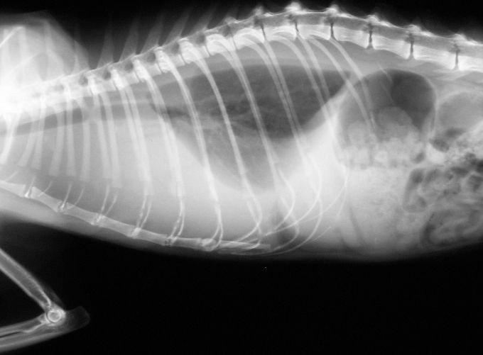
1-year-old domestic shorthair cat with fever, rapid shallow breathing and muffled heart sounds. After stabilisation with oxygen, this lateral radiograph is obtained:
 zoom_inEnlarge
Which radiographic finding is consistent with pleural effusion?
chevron_right Show answer and explanation
A. Cardiac silhouette separated from the sternum by a radiolucent band
This is the classic sign of pneumothorax on the lateral view: free air lies between the heart and sternum.
B. Thickened bronchial walls forming 'doughnuts'
This is a bronchial pattern, typical of feline asthma or bronchitis, not of pleural space disease.
C. Clearly visible outer wall of the trachea and the azygos vein
Mediastinal structures visible because they are outlined by gas indicate pneumomediastinum.
D. Interlobar fissure lines and retracted lobes with rounded margins
Correct. Fluid lies between the lobes (fissure lines) and separates them from the thoracic wall; lung margins become rounded and the cardiac silhouette and diaphragm are effaced by the silhouette sign.
lightbulb Key takeaway
In the dyspnoeic cat with suspected effusion, ultrasound (POCUS/TFAST) is more sensitive than radiography and can guide thoracocentesis before any radiograph is taken. Repeat radiographs after drainage to look for mediastinal masses, cardiomegaly or lung lesions hidden by the fluid. Heart failure is a common cause of pleural effusion in cats.
Test yourself with 10 random questions
Diagnostic imaging · 200 questions, a different test every time
Related questions
Updated: October 31, 2026 · Educational content for veterinary professionals. It does not replace current clinical guidelines or the judgement of the attending veterinarian.