Wobbler surgery with intervertebral implants: follow-up
8-year-old Dobermann with cervical spondylomyelopathy due to disc-associated compression at C5–C6 and C6–C7, treated by distraction and stabilisation with an intervertebral implant in each space. It walks with mild ataxia. The dog and its post-operative lateral cervical radiograph:
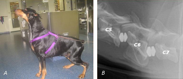 zoom_inEnlarge
Which statement about this patient's follow-up is correct?
chevron_right Show answer and explanation
A. If the follow-up radiograph is unchanged, new cord compression is ruled out
Radiographs show the implants and bone but not the cord. If signs worsen, a technique that shows the cord is needed, such as CT myelography or MRI.
B. Once these spaces have fused, compression cannot develop at other sites
On the contrary: fusion increases the load on neighbouring spaces and may promote new compression there.
C. Watch for implant subsidence or migration and compression at an adjacent space
Correct. Reported complications after distraction-stabilisation include implant subsidence into the endplates, implant migration and adjacent segment disease, which may appear months or years later and cause a relapse.
D. The intervertebral implants should be removed routinely at 3 months
Intervertebral implants are designed to stay in place and promote fusion. They are only removed or revised if they migrate, become infected or fail.
lightbulb Key takeaway
Compare each follow-up with the immediate post-operative radiograph (implant position and subsidence, fusion) and correlate it with the neurological examination. If signs recur, think of the adjacent segment and use techniques that show the cord.
Test yourself with 10 random questions
Diagnostic imaging · 200 questions, a different test every time
Related questions
Updated: October 31, 2026 · Educational content for veterinary professionals. It does not replace current clinical guidelines or the judgement of the attending veterinarian.