Tracheal collapse: radiographs, fluoroscopy and dynamic collapse
10-year-old Chihuahua with a dry “goose-honk” cough that worsens with excitement and pulling on the lead. On the lateral radiograph, taken on inspiration, the trachea has a normal diameter:
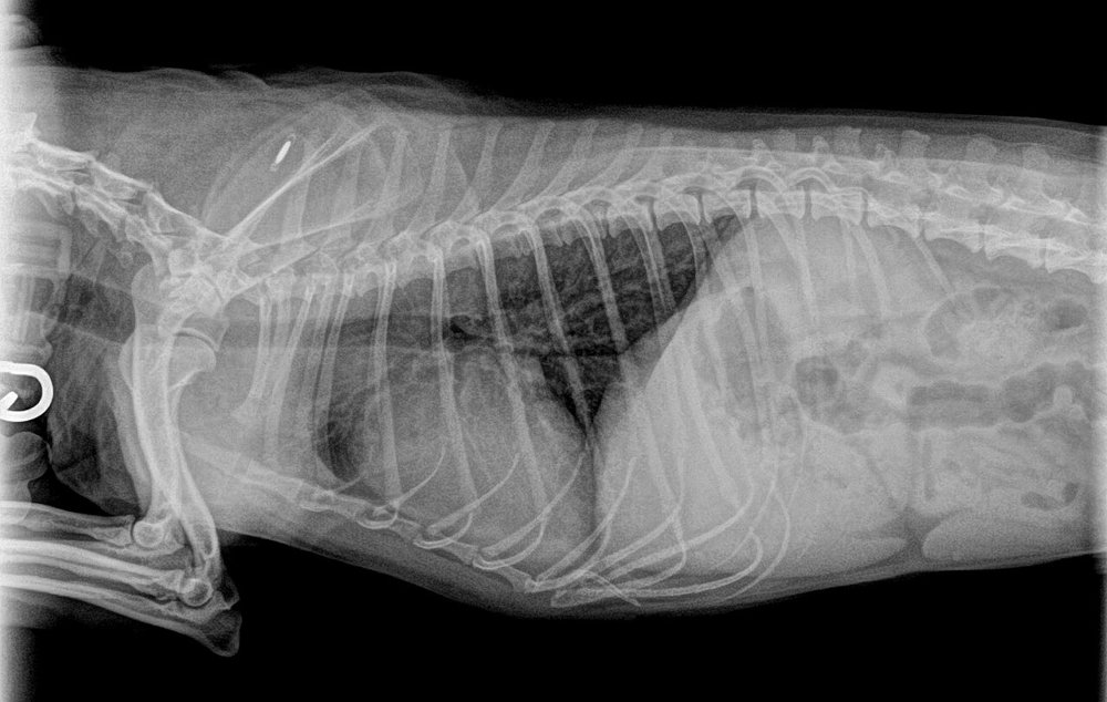 zoom_inEnlarge
Which statement is correct?
chevron_right Show answer and explanation
A. A normal tracheal radiograph rules out tracheal collapse
A static radiograph captures only one moment of the respiratory cycle and has low sensitivity; many collapses are not seen on a single image.
B. The cervical trachea collapses on inspiration and the intrathoracic trachea on expiration; fluoroscopy is more sensitive
Correct. Collapse is dynamic and depends on pressure changes. Take laterals in inspiration and expiration and, if possible, fluoroscopy (ideally while inducing a cough) or tracheobronchoscopy, which also allows grading.
C. The intrathoracic trachea collapses during inspiration
It is the other way round: during inspiration negative intrathoracic pressure tends to dilate the thoracic trachea; it collapses on expiration and especially when coughing.
D. A band of soft tissue over the trachea on the lateral always confirms collapse
Superimposition of the oesophagus or cervical muscles can mimic narrowing; this is a common radiographic false positive.
lightbulb Key takeaway
Suspect tracheal collapse in middle-aged toy breeds with a honking cough. Include the cervical trachea in the study and avoid neck hyperflexion, which can mimic collapse. Look for concurrent conditions that worsen the cough: chronic bronchitis, bronchomalacia, obesity or left atrial enlargement.
Test yourself with 10 random questions
Diagnostic imaging · 200 questions, a different test every time
Related questions
Updated: October 31, 2026 · Educational content for veterinary professionals. It does not replace current clinical guidelines or the judgement of the attending veterinarian.